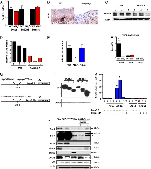
Fig. 2.
DGCR8 is a transcriptional target of ΔNp63. (A) qRT-PCR for Dicer, DGCR8, and Drosha using total RNA from epidermal cell lines of the indicated genotypes. (B) IHC using an antibody for DGCR8 on skin samples from indicated mouse embryos. (C) Western blot analysis using lysates from WT and ΔNp63−/− epidermal cell lines derived from three independent embryos (1–3) using the indicated antibodies. Actin was used as loading control. (D) Quantification of the Western blot in C. All values were normalized to WT 1. (E) qRT-PCR for DGCR8 using total RNA from epidermal cell lines of the indicated genotypes. (F) Quantitative real-time PCR of DNA purified in ChIP assay using epidermal cell lines and p63-binding site (site 1), no binding of p63 to site 2, or nonspecific binding site (NSBS). (G) Schematic showing DGCR8 site 1 (Dgcr8 S) and DGCR8 mutant of site 1 (Dgcr8 SM) luciferase reporter genes. (H) Western blot analysis using lysates from p53−/−;p63−/− MEFs transfected with the indicated p63 isoforms. Actin was used as loading control. (I) Luciferase assay for DGCR8 in p53−/−;p63−/− MEFs transfected with the indicated plasmids. Each bar represents the average of the fold activation of three independent experiments. Values are normalized to p53−/−;p63−/− MEFs transfected with vector alone. The asterisks indicate statistical significance (P < 0.001). (J) Western blot analysis of mouse embryonic stem (mES) cells, mouse-induced pluripotent stem cells (miPSYam), WT-KCs, and ΔNp63−/− epidermal cell lines expressing DGCR8 (+) or not (−) using the indicated antibodies. Asterisk indicates nonspecific band. Upper Oct4 blot is a longer exposure of the one immediately below it. Actin was used as loading control.

