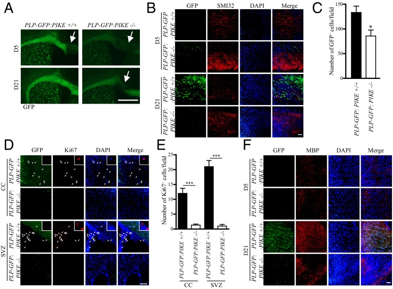
Fig. 4.
Delayed remyelination in PIKE−/− brain. (A) Representative coronal brain slices of PLP-GFP:PIKE+/+ and PLP-GFP:PIKE−/− mice (2-mo-old) collected at 5 (D5) and 21 (D21) days after LL injection. Damaged area is marked by the arrow. (Scale bar, 500 μm.) (B) SMI 32 staining in LL-injected CC after 5 (D5) and 21 (D21) days. (Scale bar, 50 μm.) (C) Number of GFP+ OLs in the damaged CC after LL injection (*P < 0.05, Student t test; n = 3). (D) Ki67 staining of the demyelinated CC in PLP-GFP:PIKE+/+ and PLP-GFP:PIKE−/− brains 5 d after LL injection. Cells that show positive Ki67 signals are indicated with arrows and are magnified in the Insets. (Scale bar, 50 μm.) (E) Number of Ki67+ cells in the CC and SVZ after LL injection (***P < 0.05, Student t test; n = 3). (F) IF staining of MBP in the LL-damaged CC. (Scale bar, 50 μm.)

