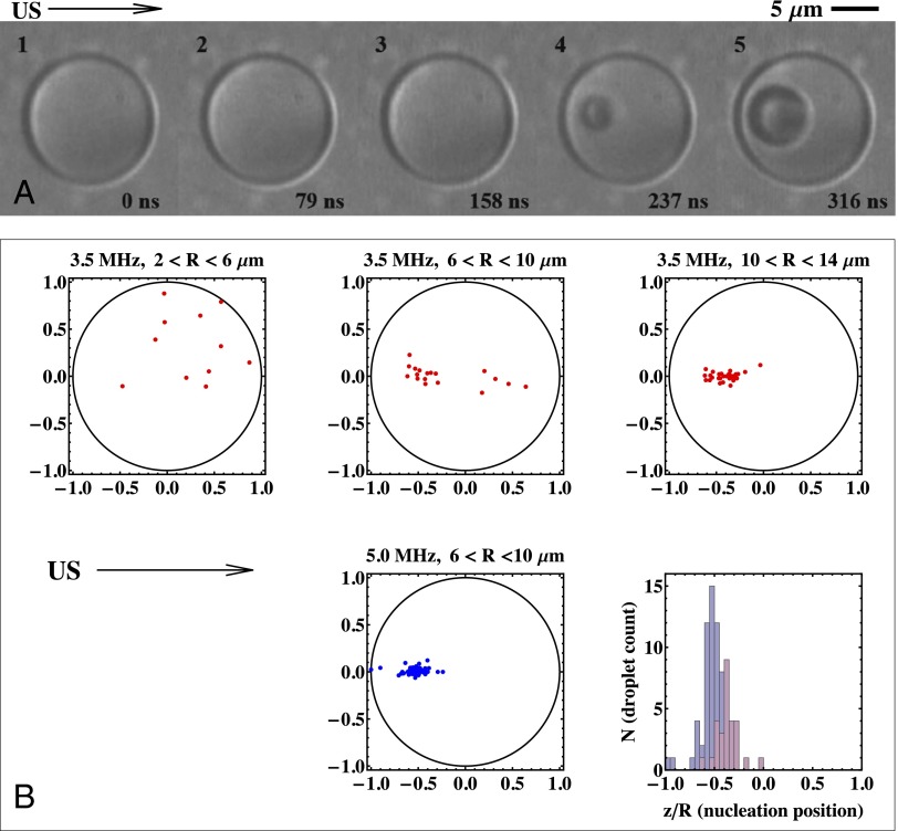
Fig. 5.
(A) A set of consecutive images showing acoustic droplet vaporization of a 7.4-μm-radius PFP droplet taken at a frame rate of 12.6 million frames per second. The droplet is triggered by an eight-cycle, 5-MHz frequency ultrasound pulse. The nucleation is initiated between frames 3 and 4. Frames 4 and 5 show the subsequent vapor bubble growth (24). (B) Nucleation maps for the two frequencies and for a range of droplet sizes. The histogram shows the focused positions z/R for a frequency of 3.5 MHz for the 10- to 14-μm droplet sizes and for a frequency of 5.0 MHz for the 6- to 10-μm droplet sizes. US, ultrasound.

