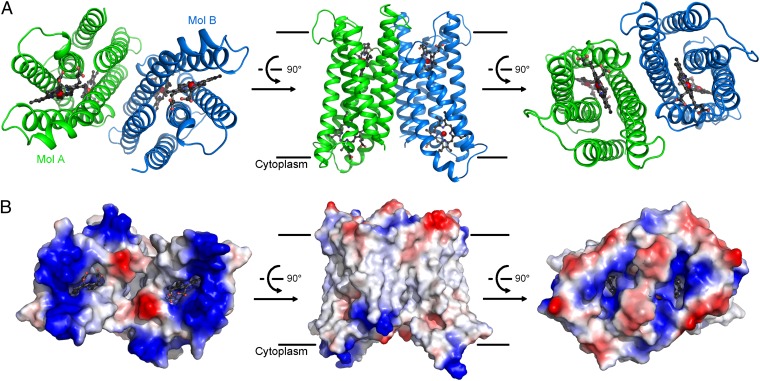
Fig. 1.
Overall structure of Cyt b561. (A) Overall structure of WT, full-length Cyt b561. The structure of Cyt b561 is shown in three successive views. There are two molecules of Cyt b561 in each asymmetrical unit, named Mol A (green) and Mol B (blue). (B) Surface features of the Cyt b561 homodimer by electrostatic potential. The three views shown correspond to those in A. Two cavities on either side are surrounded by positively charged amino acids. All structural figures were prepared using PyMOL Molecular Graphics System, Version 1.5 (Schrödinger, LLC).

