
Fig. 1.
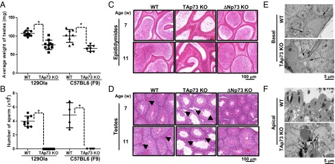
Testes of TAp73 KO mice exhibit altered germ cell numbers, germ cell localization, testicular histology, and ultrastructure. (A and B) The average weights of testes (A) and the number of mature sperm isolated from epididymides (B) were measured in 36-wk-old WT (n = 10) and TAp73 KO (n = 10) mice of the 129Ola background and in 16-wk-old WT (n = 8) and TAp73 KO (n = 8) mice of the C57BL6 (F9) background. Data points are values for individual mice. The horizontal line is the group mean ± SD (*P < 0.02; unpaired Student t test). (C and D) H&E-stained histological sections of epididymides (C) and testes (D) of WT, TAp73 KO [C57BL6 (F9)], and ΔNp73 KO (control) mice of 7 or 11 wk of age. Arrowheads show Leydig cells. (E and F) Electron micrographs of testes from 36-wk-old WT and TAp73 KO mice. (E) Normal basal ES (Basal) of BTB (arrows) surrounding the lower lateral membranes of Sertoli cells in WT and TAp73 KO mice. (F) In contrast to orderly apical ES (apical) in WT testes, TAp73 KO testes show disorganized apical ES populated by malformed spermatids (arrows).