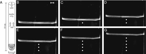
Fig. 2.

Thiovulum majus cells aggregate into a front that propagates up the oxygen gradient. (A) A schematic of the experiment. Bacteria are inoculated above a sulfide source at the base of a test tube. As cells consume oxygen, the front moves up the test tube. (B–G) Images of the front at 15-min intervals. The front is shown in profile. White dots show the position of the front in the preceding stills. (Scale bar, 1 mm.)
