Fig. 2.
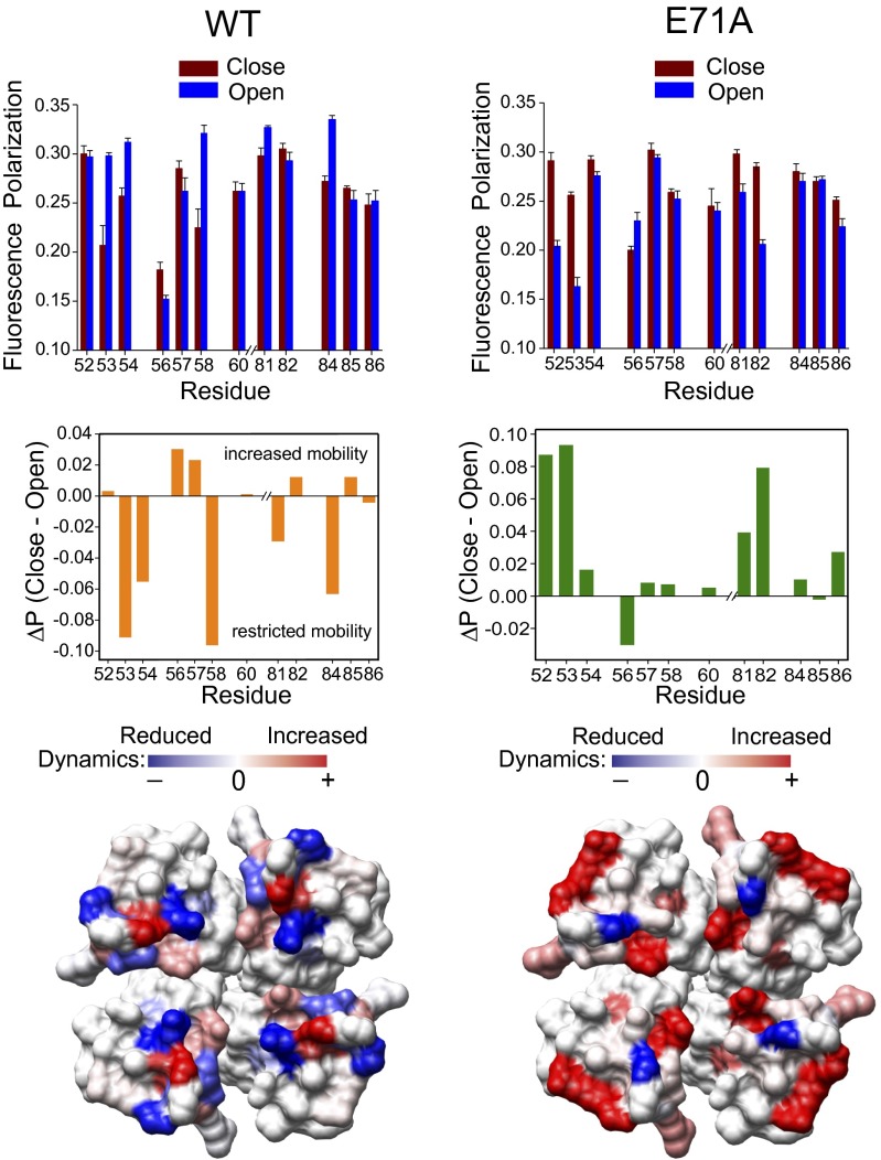
Outer vestibule of the open/noninactivating conformation is highly dynamic. (Top) Steady-state polarization of NBD fluorescence measured for NBD-labeled outer vestibule residues of full-length WT (Left) and E71A (Right) KcsA reconstituted in POPC/POPG (3:1, moles/moles) liposomes. Measurements at pH 7.5 and pH 4 correspond to the closed and open states of the channel, respectively. The excitation wavelength used was 480 nm; emission was monitored at 535 nm in all cases. The difference in polarization values (ΔP) represent mean ± SE of three independent measurements. The ΔPs (Middle) are shown and were mapped on the crystal structure of KcsA (1K4C) to highlight the changes in dynamics between the different functional states of KcsA (Bottom) upon gating. Details are provided in SI Materials and Methods.

