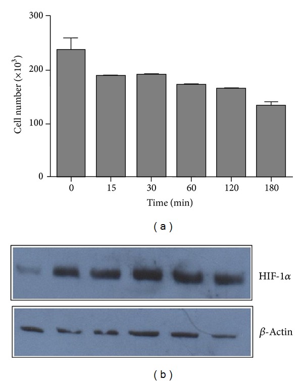
Figure 1.

Induction of cell death by the coverslip hypoxia model. (a) Coverslips were placed onto confluent cardiac myocyte monolayers and removed after 15, 30, 60, 120, and 180 min. As a negative control, myocyte cultures were left uncovered. Cell viability was evaluated by the MTT assay. Data represent the average of three independent tests. Error bars indicate the standard error of the mean. (b) Proteins in whole cell lysates obtained at the indicated time points were analyzed for HIF-1α expression by immunoblotting. As a control, the expression of the constitutive β-actin protein was evaluated.
