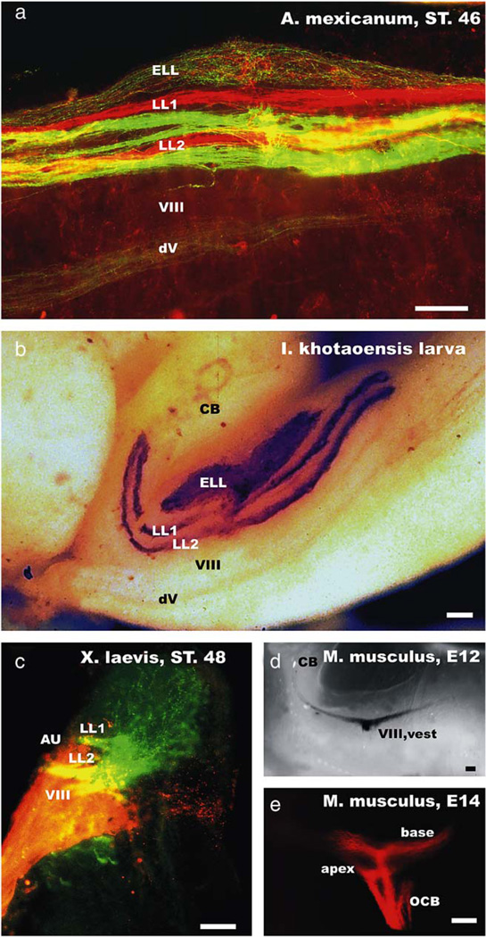
Fig. 1.
The organization of the lateral line and inner ear afferent projections are shown for the axolotl (a), a gymnophionan (b), a frog (c) and the vestibular and cochlear projections of a mouse embryo (d, e). Note that older salamander and gymnophionan larvae show a clear segregation of mechanosensory afferents of a given peripheral lateral line nerve into two distinct fascicles (LL1, LL2). Labeling two peripheral nerve branches (in this case the anterodorsal and anteroventral lateral line nerve) with differently colored dextran amines results in labeling of two discrete fascicles for each nerve (a). In contrast, no such fasciculation is apparent in the electrosensory afferent projections (ELL; a, b). Frog lateral line projections show two entering fascicles, but widespread ramification throughout the entire lateral line nucleus (c). The frog inner ear projection shows the vestibular component (VIII) ventral to the lateral line projection, but also the formation of the auditory projection (AU) lateral to the lateral line projection. In mice, the vestibular (d) and cochlear (e) fibers are forming distinct projections from the earliest time they can be labeled and do so even for subdivisions derived from different parts of the cochlea (e). Labeling of all cochlear afferent fibers would result in a continuous projection with no distinct fascicles being apparent. CB, cerebellum; dV, descending tract of the trigeminal nerve; LL1, LL2, lateral line afferent fascicles; ELL, electroreceptive ampullary organ projection; VIII, vest, vestibular component; OCB, olivocochlear bundle. Bars indicate 100 µm in all images.

