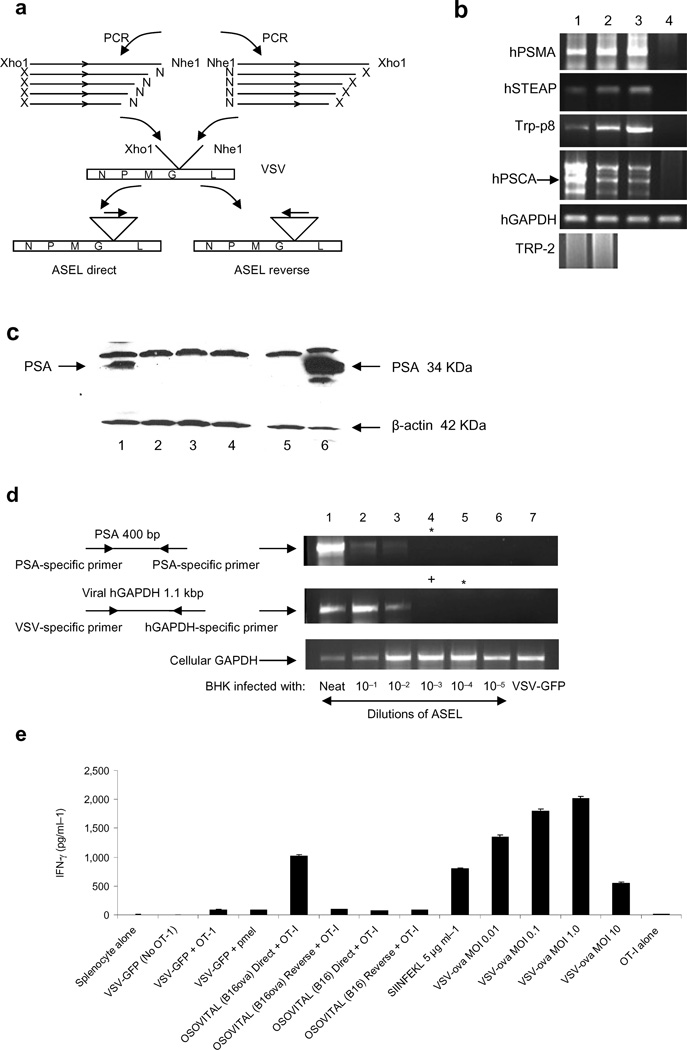
Figure 1. VSV-expressed cDNA libraries.
a. The ASEL VSV-expressed cDNA library contains cDNA from normal human prostate cloned into VSV in direct, or reverse, orientation. b. Human prostate specific genes37–40, but not the melanocyte-specific TRP-211, detected by PCR in the original human prostate plasmid library (lane 1) and in the VSV-cDNA plasmid library (2). rtPCR from cDNA of HT1080 cells infected with ASEL (MOI 0.1) (3) compared to uninfected cells (4) (predicted size for hPSCA shown with an arrow). c. BHK cells infected with ASEL Direct (1) or Reverse (2), or with control viruses (lanes 3&4) (MOI ~10) assayed for human PSA by Western Blot. Lane 5, uninfected BHK cells; Lane 6, 104 human prostate LnCap cells. d. BHK cells infected with 10 fold dilutions of ASEL virus (1–6) assayed by rtPCR for PSA or human GAPDH. No PSA-specific signal was detected at dilutions lower than 1:100 of the original virus stock. (Expression of GFP from 100 pfu of VSV-GFP could be detected by this assay). +Positive/*negative for PCR upon nested PCR. e. Splenocytes infected with VSV-GFP or VSV-cDNA libraries from cells which did, or did not, express OVA (MOI 0.1), co-cultured with naive OT-I T cells and assayed for IFN-γ28. Lane 1, splenocytes alone; (2–4), splenocytes infected with VSV-GFP, without OT-I (2), with OT-I (3) or with irrelevant T cells41; (5&6), splenocytes infected with VSV-cDNA library from B16ova cells in Direct (5) or Reverse (6) orientation with OT-I; (7&8), splenocytes infected with VSV-cDNA library from B16 cells (no OVA) in Direct (7) or Reverse (8) orientations with OT-I. (9), OT-I activated by SIINFEKL peptide. (10–13), splenocytes infected with VSV-ova at MOI 0.01 (10), 0.1 (11), 1.0 (12) and 10 (13). (14), OT-I (no splenocytes, no VSV).

