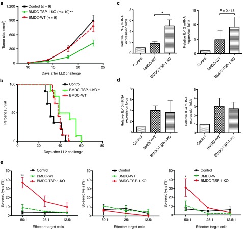
Figure 4.
TSP-1–deficient dendritic cells displayed stronger antitumor effects than control dendritic cells. (a) TSP-1–deficient dendritic cells enhanced anticancer therapeutic effects. LL2 tumor-bearing mice were injected subcutaneously with 1 × 10⁵ bone marrow–derived dendritic cells (BMDCs) isolated from TSP-1 knockout (KO) or wild-type (WT) mice. The tumor growth was measured (**P < 0.01, BMDC-TSP-1 KO versus BMDC-WT). (b) TSP-1–deficient DCs prolong the survival of mice. Kaplan–Meier analysis was performed on the mice survival data (*P < 0.05, BMDC-TSP-1 KO versus BMDC-WT). (c) Bar graphs represent the quantification of Th1 cytokines expression in the lymph nodes. (d) Th2 cytokines expression in the lymph nodes. Lymph node messenger RNA (mRNA) was isolated after immunization. Interferon-γ (IFN-γ), interleukin (IL)-12, IL-4, IL-10, and HPRT mRNA expression levels were measured by real-time polymerase chain reaction The relative expression fold was compared with control group. *P < 0.05, BMDC-TSP-1 KO versus BMDC-WT; n = 4 mice per group, mean ± SEM. (e) Splenocytes from mice that received TSP-1-KO BMDCs were cytotoxic against tumor cells. Effector cells were isolated from mice that received the indicated treatments. LL2 luciferase cells were used as target cells. Left panel: cytotoxicity splenocytes against LL2 tumor cells. Middle panel: cytotoxicity of CD8+ T-cell–depleted splenocytes against LL2 tumor cells. Right panel: cytotoxicity of NK-cell–depleted splenocytes against LL2 tumor cells (**P < 0.01, BMDCs-TSP-1 KO versus BMDCs-WT; *P < 0.05, BMDCs-TSP-1 KO versus BMDCs-WT). shRNA, short hairpin RNA; TSP-1, thrombospondin 1.

