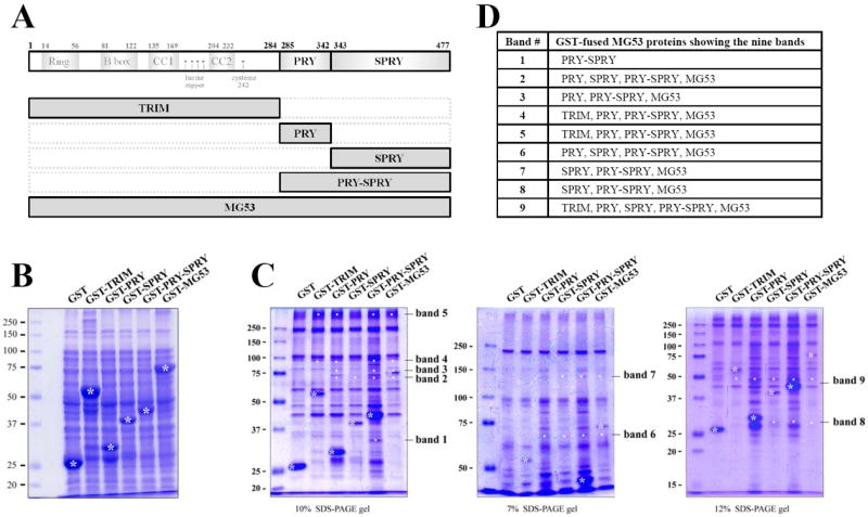
Figure 1. Binding assays of GST-fused MG53 proteins with triad proteins.

(A) Schematic diagrams of full-length mouse MG53 and domains. Numbers indicate the sequence of amino acids. (B) GST-fused MG53 proteins expressed in E.coli were separated on a SDS-PAGE gel (10%) and stained with Coomassie Brilliant Blue staining. GST-fused MG53 proteins are indicated by white asterisks. (C) The bound proteins obtained from the binding assays of GST-fused MG53 proteins with the triad proteins from rabbit skeletal muscle were separated on three different percentages of SDS-PAGE gels and stained with Coomassie Brilliant Blue. GST was used as a negative control. GST or GST-fused MG53 proteins are indicated by white asterisks. The specifically bound proteins to the GST-fused MG53 proteins are indicated by white dots. The newly appearing nine bands compared with the GST control are indicated on the right (bands 1 to 9). (D) The GST-fused MG53 proteins showing the nine bands are summarized.
