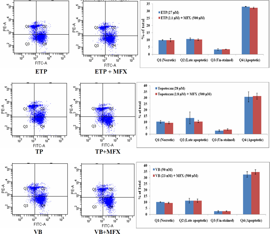
Figure 6.
Flow cytometry analysis using Annexin V (AV) and propidium iodide (PI) staining illustrating modulation of etoposide (ETP), topotecan (TP), and vinblastine (VB) mediated Y-79 cell apoptosis by moxifloxacin (MFX). Q1= PI positive cells (AV−PI+); Q2 = Late apoptotic (dead) cells (AV+PI+); Q3 = Un-stained (non-apoptotic healthy) cells (AV−PI−); Q4 = Early apoptotic (but viable) cells (AV+PI−).

