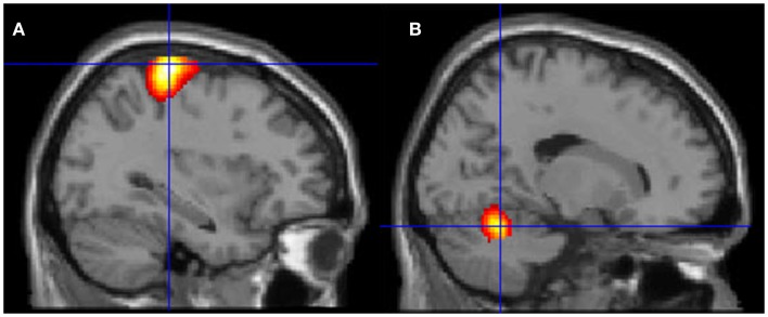
Figure 6.
Motor areas significantly activated more during movement of her fingers to thumb compared with visualizing the same movement. (A) Representation of the left primary motor cortex; (B) representation of the right cerebellum. The p-value for this image was set to 0.001 uncorrected with the cluster threshold at 100 significant voxels.

