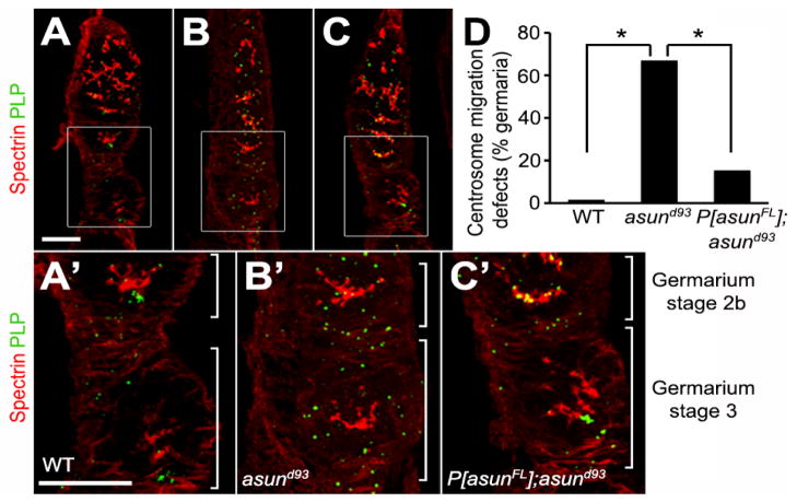
Figure 7. Centrosome migration defects of asund93 germaria.
(A–C) Projections of confocal sections of wild-type, asund93, and rescued asund93 germaria stained for spectrin (red; fusome marker) and PLP (green; centriole marker). Enlarged insets shown below. Anterior, top; dorsal, left. Within wild-type cysts, most centrosomes have migrated from the nurse cells into the oocyte (located at posterior of egg chamber) by stage 2b of the germarium (A, A′). Centrosomes do not properly migrate into the oocyte in asund93 germaria and are found distributed throughout the entire cyst (B, B′). Centrosome migration occurs in rescued germaria but is delayed (C, C′). Scale bars, 20 μm. (D) Quantification of centrosome migration defects in wild-type, asund93, and rescued asund93 germaria (>100 germaria scored per genotype). Asterisks, p<0.0001.

