Figure 2.
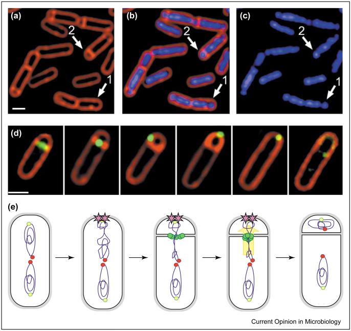
Chromosome segregation during B. subtilis sporulation. (a-c) Micrograph showing B. subtilis cells early in sporulation, following staining with FM 4-64 to visualize cell membranes (red) and DAPI to show DNA (blue). Prior to polar septation, the origin-proximal 30% of the future forespore chromosome condenses near one cell pole (arrow 1); the asymmetrically-positioned sporulation septum is synthesized between these two chromosome domains (arrow 2). (d) Localization of the SpoIIIE DNA translocase (green) together with cell membranes (red). The protein first forms a ring at the site of cell division, then assembles a focus at the septal midpoint, and finally it relocalizes to the cell pole, where it participates in the final stages of the phagocytosis-like process of engulfment [82]. Scale bars = 1 μm. (e) Model for chromosome segregation during sporulation. At the onset of sporulation, the origins (yellow-green circles) are brought to the cell poles by an interaction with RacA (purple stars), while the temini remain near mid-cell (red circles). During septation, the SpoIIIE DNA translocase (green circles) assembles at the septal midpoint, and translocates the forespore chromosome across the septum (yellow arrow).

