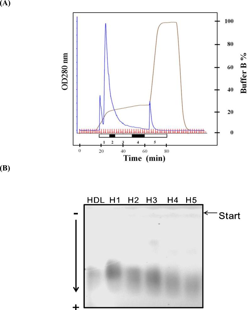
Figure 1.
Separation of HDL into 5 distinct subfractions according to charge. (A) FPLC elution profiles of the 5 HDL subfractions separated by using a UnoQ12 column and the Tris-HCl buffer system at a flow rate of 2 mL/minute. Approximately 100 mg of HDL in 10 mL was loaded onto the column and separated with a multistep gradient of buffer B. Elution was monitored at 280 nm. (B) Results of agarose gel electrophoresis of HDL and subfractions H1 through H5. Each HDL sample (2.5 μg in 9 μL) was loaded onto a 0.7% agarose gel and separated at 100 V for 1.4 hours.

