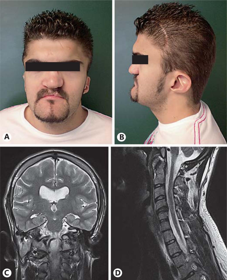
Fig. 1.
Anterior (A) and lateral (B) views of the patient's face showing normal hair, broad forehead, short neck, broad nasal base with repaired cleft lip and palate, normal eyebrows and eyelashes, bilateral microtia with low-set ears, hypoplastic lobule, narrow helix, prominent antihelix, and a triangular concha. It also shows a scar secondary to a previous craniectomy. C Cerebral magnetic resonance imaging coronal view revealing moderate dilatation of the ventricular system without hydrocephalus and mild cerebral atrophy. D Cerebral magnetic resonance imaging showing intervertebral osteochondrosis of C4-C5, C5-C6 and C6-C7, and C4-C5 with discrete right neural foramen stenosis.

