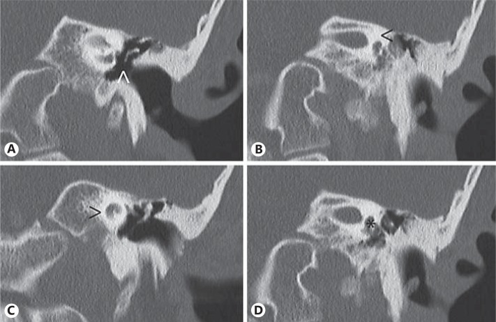
Fig. 2.
Left ear computed tomography showing partial anvil and total stirrup aplasia (arrowhead in A), complete absence of the semicircular canals (arrowhead in B), bilateral hypoplasia of the cochlea (arrowhead in C) with a diminishment in size of the basal and apical turns, and dysplasia of the vestibule (asterisk in D).

