
Figure 5.
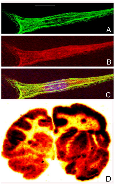
Panels A, B, and C are confocal micrographs of MLD fibroblasts incubated with the HIRMAb-ASA fusion protein (10 ug/mL) for 24 hours. The cells were fixed with 100% methanol and immune stained for confocal microscopy. The fixed cells were stained with a mouse monoclonal antibody to human lysosomal associated membrane protein (LAMP)-1 (panel A: green channel) and a goat polyclonal antibody to human ASA (panel B: red channel). The overlap image in panel C shows sequestration of the HIRMAb-ASA fusion protein within lysosomes. The magnification bar in panel A is 15 microns. Panel D is film autoradiography of a coronal section of Rhesus monkey brain removed 2 hours after the IV administration of the [125I]-HIRMAb-ASA fusion protein. The study shows global distribution of the fusion protein throughout the brain with higher uptake in gray matter as compared to white matter.