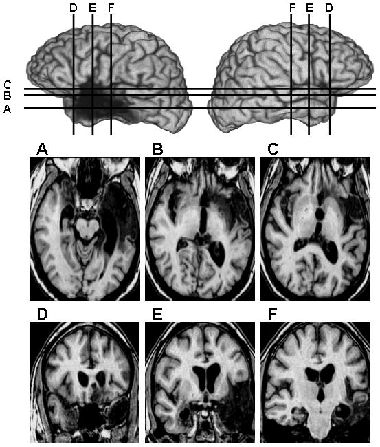
Figure 1. MRI scans of patient SZ’s brain.

Lateral views of the left hemisphere (left) and right hemisphere (right) are shown in the upper section of the figure, and axial (A–C, middle row) and coronal (D–E, bottom row) slices are shown below. (A) Axial slice depicting bilateral damage to the medial temporal lobe, medial temporal poles, and unilateral damage to a large region of the left temporal lobe. (B & C) Axial slices depicting bilateral damage to the insular cortex and left-sided damage to the basal forebrain and posterior orbitofrontal cortex. (D) Coronal slice depicting bilateral damage to the temporal poles (with only the medial temporal pole affected on the right side), and unilateral damage in the region of the left basal forebrain. (E) Coronal slice depicting bilateral damage to the amygdala and insula, and unilateral damage to a large region of the left temporal lobe. (F) Coronal slice depicting bilateral hippocampal damage and some damage to the left temporal cortices and left posterior insula. All images use radiological convention (i.e., right side of image = left hemisphere and vice versa).
