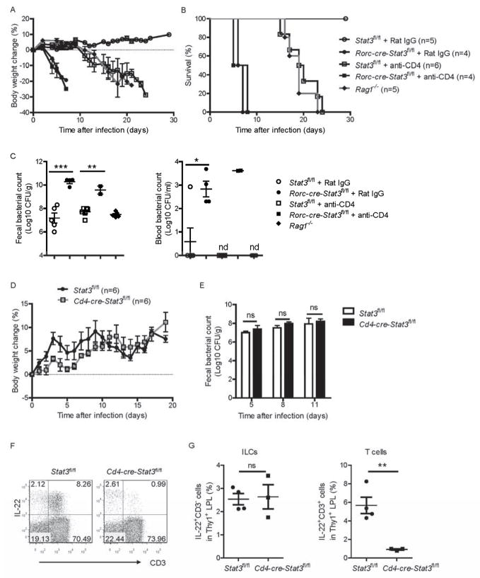
Figure 4. STAT3 signaling and IL-22 from RORγt+ ILCs, but not T cells, are essential for the protection from C. rodentium infection.
(A–C) Rorc-cre-Stat3fl/fl and wild type Stat3fl/fl mice were infected with 5 × 10⁶ CFU of C. rodentium and injected intraperitoneally with mAb GK1.5 or Rat IgG (50 μg per mouse each time) at day −5, 0, 5, 10 and 15 post infection for depletion of CD4+ T cells (4 to 6 mice per group). Average body weight change (A) and survival rates (B) at the indicated time points are shown. (C) Bacterial titers from blood and fecal homogenate cultures at day 5 post infection. Each dot represents one individual mouse.
(D, E) Cd4-cre-Stat3fl/fl (n=6) and wild type Stat3fl/fl mice (n=6) were orally infected with 2 × 10⁹ CFU of C. rodentium. (D) Body weight changes are shown at the indicated time points. (E) Bacterial titers from fecal homogenate cultures at indicated day post infection in the mice in D.
(F, G) IL-22 expression in CD3− and CD3+ cells were analyzed by intracellular cytokine staining followed by flow cytometry. Cd4-cre-Stat3fl/fl and wild type Stat3fl/fl littermate mice were infected with 2 × 10⁹ CFU of C. rodentium. LPLs were isolated from the colons at day 8 post-infection. Cells were stimulated with IL-23 (25 ng/ml), PMA (50 ng/ml) and ionomycin (750 ng/ml) for 4 hours and gated in Thy1+ lymphocytes. Each dot represents one individual mouse.
*P<0.05, **P<0.01, ***P<0.001; ns, no significant difference (Student’s t-test). nd, nondetectable. Data are representative of two independent experiments (A–G; mean ± s.e.m.). See also Figure S4.

