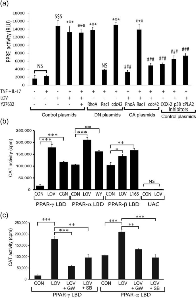
FIGURE 5.
LOV enhances PPARs transactivation in B12 cells via production of PPARs ligands. B12 cells (2,000 × cm2) were treated with LOV (2.0 μM) or other pharmacological agents after transient transfection with the plasmids for PPRE and β-galactosidase activity or with PPAR-GAL4/(UAS)5-CAT reporter system. (A) The composite mean ± SE of three to four samples analyzed in triplicate depicts PPRE reporter activity/β-galactosidase activity in treated B12 cells for 24 h after transfection with control/or DN/CA forms of the RhoA, cdc42, and Rac1 plasmids and/or treated with inhibitors of COX-2 (0.25 μM), p38 MAPK (SB203580; 2 μM), and cPAL2 (2 μM). (B and C) The composite mean ± SE of three to four samples analyzed in triplicate depict CAT reporter activity in treated B12 cells for 24 h. Data are presented as counts per minute (cpm) normalized to β-galactosidase activity. Pharmacological agents were agonists of PPAR-γ (ciglitazone; 0.5 μM), PPAR-α (WY14643; 100 μM), and PPAR-β (L-165041; 20 nM) or inhibitors of PPARγ(GW9662; 0.25 μM), PPAR-α (GW6471; 0.1 μM), and p38 MAPK (SB203580; 2 μM). Statistical significance as indicated in Fig. 1 legend for B and C. For A, ***P < 0.001 and NS (not significant) versus TNF-α and IL-17; ###P < 0.001 versus TNF + IL-17 + LOV or Y27632 and $$$P < 0.001 versus no treatment. RLU; relative light units.

