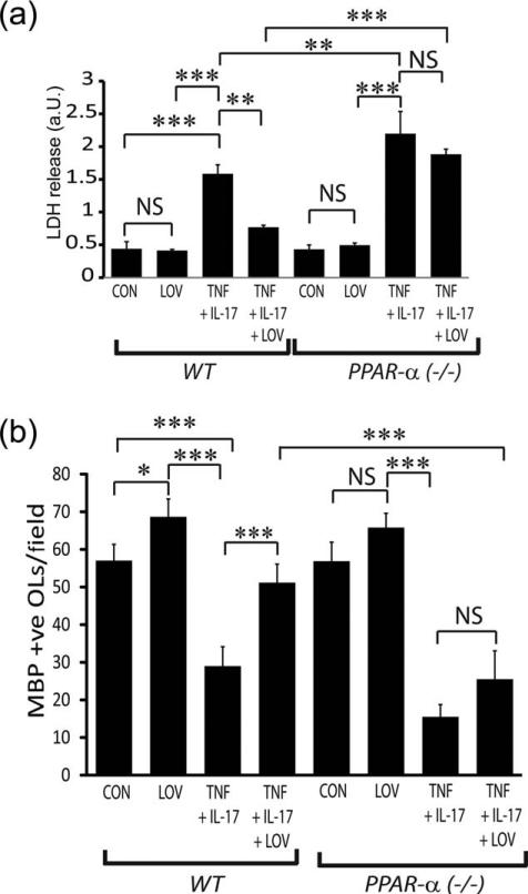
FIGURE 8.
LOV-mediated protective effects were lacking in PPAR-α-deficient OLs exposed to cytokines. OLs from wild type (WT) and PPAR-α(–/–) mice were exposed to TNF plus IL-17 and LOV as detailed in the Fig. 1 legend. (A) The composite mean ± SE of three to four samples analyzed in triplicate depicts LDH release in the culture supernatants of treated OLs for 96 h. (B) The composite mean ± SE of three to four slides analyzed in triplicate depicts a MBP+ve OLs/field of the slide in treated OLs for 96 h. Statistical significance as indicated in Fig. 1 legend.

