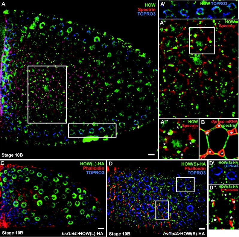
Figure 3.
HOW(S) localizes near the basal plasma membrane of follicle cells at stage 10 of oogenesis. A–A″′. Immunolocalization of endogenous HOW using an anti-HOW antibody (green) in stage 10 Drosophila follicular epithelium. α-spectrin marks the cell cortex (red) and TOPRO3 marks the nucleus (blue). HOW is localized in the nucleus (see top insert, A′) as well as in the cytoplasm (A″), in particular in dots around the open ZOC (asterisks in insert bottom, A″′) at the basal side of the follicle cells. Note that the nucleolus (unstained dot in the middle of the nucleus is very large). (B) FISH localization of dgrasp mRNA (red) with respect to the open ZOC (asterisks). α-spectrin (green) marks the cell cortex. (C) Immuno-localization of HOW(L)-HA overexpressed in the follicular epithelium using the UAS-Gal4 system under the control of a heat-shock promoter. Anti-HA labelling (green) shows that HOW(L) is restricted to the nucleus at all stages in the follicular epithelium development. Phalloidin (red) stains the actin of the cell cortex and TO-PRO®-3 (blue) marks the nucleus. (D–D″) Immunolocalization of HOW(S)-HA (using an anti-HA antibody, green) overexpressed in the follicular epithelium using the UAS-Gal4 system under the control of a heat shock promoter, Phalloidin (red) stains the actin of the cell cortex and TO-PRO®-3 (blue) marks the nucleus. Inserts show that HOW(S) is absent from the nucleus (D′) whereas it is enriched around the open ZOC (asterisks, bottom insert). Scale bars: 10 µm.

