
Figure 1.
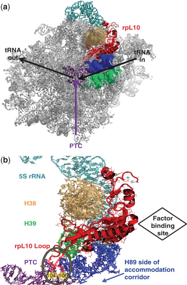
rpL10 is strategically positioned in the core of the LSU. (a) The big picture: rpL10 in the context of the subunit interface of the LSU. (b) A close-up view of rpL10 and the local environment. The hypothetical loop structure is circled and indicated by dashed red lines, and the approximate positions of S104 and A106 are indicated. The protein is situated between Helices 38 and 89, and appears to be an extension of Helix 39. It is located in close proximity to several functional centers of the LSU including the PTC, the aa-tRNA AC and the elongation factor binding site. It is also positioned to communicate with the SSU through Helix 38 and the 5S rRNA. Images were generated using PyMOL.