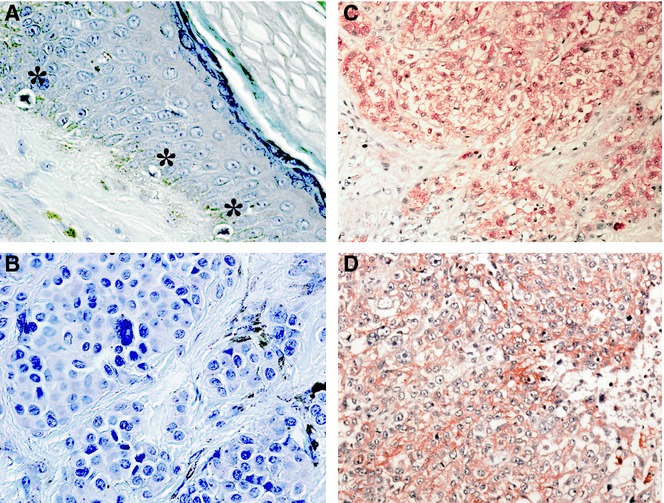
Figure 1.

Overexpression of Nedd4L in melanoma tissues. The immunohistochemical staining showed that the immunoreactivity of Nedd4L was minimal or none in non-tumourous melanocytes or in benign nevus cells. In contrast, immunoreactivity of Nedd4L was observed in the cytoplasm of 34 of 79 cutaneous melanomas and 9 of 32 nodal metastatic melanomas. The figure shows representative stainings. (A) Non-tumourous melanocytes indicated by asterisk (*) were not stained. (B) No significant Nedd4L immunoreactivity was observed in benign nevus. (C and D) Representative Nedd4L staining in cutaneous melanoma (C) and nodal metastatic melanoma (D).
