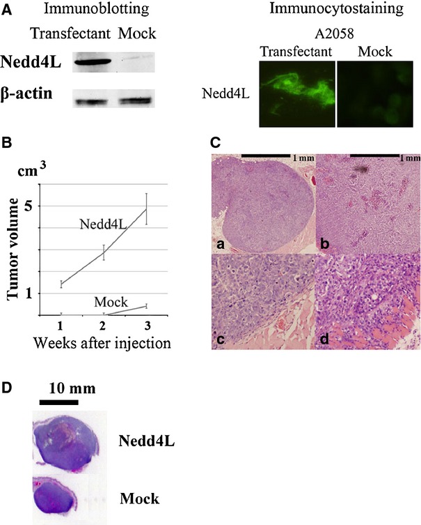
Figure 3.

Nedd4L expression accelerated melanoma cell growth in vivo. (A) Representative data of immunoblotting and immunocytostaining of A2058 melanoma cells showing the exogenous expression of Nedd4L. Original A2058 cells expressed less amount of Nedd4L protein (Figure ;2A). Clones of A2058 cells exogenously expressing Nedd4L and control cells not expressing Nedd4L were established by transfection with an expression vector, which contained the entire coding region of Nedd4L cDNA, or a vector alone. (A) representative result from three independent immunoblotting experiments using different transfectant clones is shown. (B) We subcutaneously injected 5 ;× ;105 cells into nude mice. Nedd4L-expressing A2058 cells formed a visible tumour in all the five transplanted BALB/c nude mice after 1 ;week of inoculation (indicated as Nedd4L). By contrast, control A2058 cells formed a visible tumour in none of the five transplanted BALB/c nude mice after 1 ;week or 2 ;weeks of inoculation (indicated as Mock). The tumour volume of each mouse was calculated every week using the formula: volume ;= ;(d1 ;× ;d2 ;× ;d3) ;× ;0.5236, where dn represents the three orthogonal diameter measurements. Representative data are demonstrated as mean ;± ;standard error of the mean (SEM). Similar results were obtained from another independent experiment using different transfectants. (C) Representative histological features of xenograft specimens. In total, 5 ;× ;105 cells were subcutaneously injected into nude mice. Haematoxylin-eosin-stained tissue specimens of transplanted tumours harbouring non-Nedd4L-expressing A2058 cells (a and c) and Nedd4L-expressing A2058 cells (b and d) at 3 ;weeks are shown. Note the rounded tumour border of the non-Nedd4L-expressing A2058 cells with a thin fibrous capsule (c). By contrast, Nedd4L-expressing A2058 tumour exhibited deep invasion with destruction of the muscle layer (d). (D) Whole tumour silhouette of the representative transplanted Nedd4L-expressing A2058 cells (indicated as Nedd4L) and control non-Nedd4L-expressing cells (indicated as Mock) after 3 ;weeks of inoculation to compare the tumour size at glance.
