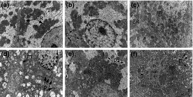
Figure 2.

Electron micrographs of liver tissues in the study groups. Uranyl acetate–lead citrate, a, b, c, d, e, f ×12000. a: Control; b: SeS; c: SeD; d: di(2-ethylhexyl)phthalate (DEHP); e: DEHP/SeS; f: DEHP/SeD groups. Nucleus (n), endoplasmic reticulum (ER), mitochondria (m), peroxisome (p) were marked. Normal ultrastructural features of hepatocytes with the normal arrangement of ER and mitochondria are observed in C and SeS groups (a and b). The glycogen granules are prominent in the cytoplasm of SeS group (c). The cytoplasm is filled with damaged mitochondria in DEHP group (d). The crista of mitochondria cannot be observed in DEPH/SeD group (e).
