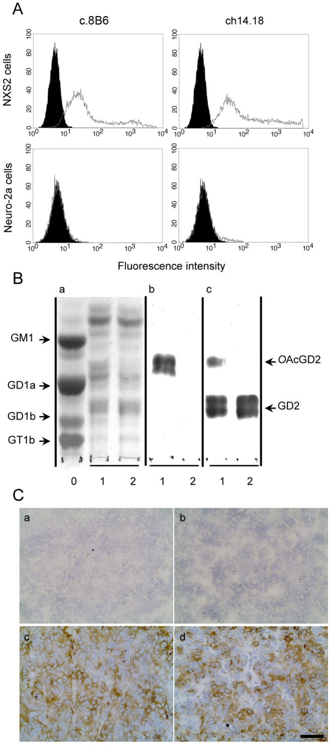
Figure 1. Binding specificity chimeric mAbs c.8B6 and ch14.18 measured by flow-cytometric analysis (A), by TLC-immunostaining (B) and by Immunochemistry on NXS2 tumor (C).

(A) NXS2 and the OAcGD2/GD2− Neuro2A cells were stained as described in Materials and Methods. Representative flow cytometry histogram of OAcGD2 expression. Antibody c.8B6 or mAb ch14.18 (grey) and control antibody (black). The same staining pattern was observed for mAb c.8B6 or ch14.18 on OAcGD2/GD2+ NXS2 cells whereas neither mAb c.8B6 nor mAb ch14.18 bind OAcGD2/GD2-negative Neuro-2a cells. These experiments were independently replicated 3 times. (B) TLC of gangliosides extracted from rat brain (Lane 0) and NXS2 cells with (Lane 1) or without (Lane 2) alkaline treatment and stained with resorcinol-HCl (Panel a) or immunostained with mAbs c.8B6 (Panel b), or ch14.18 (Panel c). Chimeric mAb 8B6 reacted with the alkali-labile OAcGD2 ganglioside with no cross-reactivity against GD2 ganglioside (Panel b) whereas ch14.18 reacted with GD2 and OAcGD2 (Panel c). The same results were obtained in 3 independent experiments. (C) An immunoperoxidase assay was performed on NX2 neuroblastoma tumor sections as described in Material and Methods. Strong immunostaining was detected on neuroblastoma cells with either mAb c.8B6 (c) or mAb ch14.18 (d). The anti-CD20 chimeric antibody ritximab was used as a negative control (b). No antibody (a). NXS2 neuroblastoma tumors from 6 different mice were tested with the same results. Scale bar = 20 µm.
