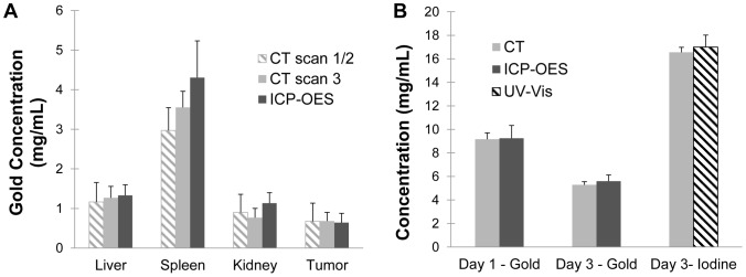
Figure 10. Comparison of CT-measured concentrations with validation methods.

(A) Concentrations of accumulated gold in the tumors and other organs for the single-material method (based on CT scan 1 and 2), the two-material method (based only on CT scan 3) and ICP-OES. (B) Measured CT concentrations of gold in the blood on day 1 and day 3, iodine concentration in the blood on day 3, and corresponding measurements by ICP-OES and UV-Vis. None of the differences were found to be statistically significant (p<0.05).
