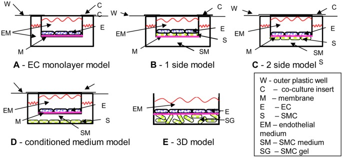
Figure 1. The major vascular wall models found in the literature.
A - EC monolayer model, presented on a membrane but can be on the outer plastic as well. B – 1-side model, where EC are cultured on a confluent culture of SMC. C – 2-side model, where EC and SMC are cultured on opposite sides of a porous membrane. The cell can form connections and usually shares the same medium. D – Conditioned medium model, where the cells share the same medium, but no connections can be formed between the cells, and E – EC are cultured on a polymerized gel containing SMC, usually collagen gel.

