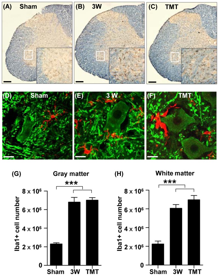
Figure 5. Microglia markedly increased in number in the lumbar motor region following thoracic SCI.

(A-C) Representative images of lumbar spinal cord sections stained with an antibody recognizing the microglial marker Iba1 (dark brown) antibody in sham operation (A), at 3 weeks (3W) after injury (B), and at 3W after injury with treadmill training (TMT) (C). Immunostained sections were counterstained with eriochrome cyanine to differentiate the gray matter and white matters. Insets are magnified images of the regions in the white boxes in the ventral gray matter. Scale bars represent 100 µm. (D-F) Representative images of lumbar spinal cord sections colabeled with Iba1 (red) and MAP2 (green) antibodies in sham operation (D), at 3W after injury (E), and at 3W after injury with TMT (F). Note that Iba1 positive microglial cells are frequently associated with MAP2 positive neurons and dendritic neuropil areas. Scale bars represent 20 µm. (G-H) Quantification of stereological counting of Iba1-positive microglial cells in the gray matter (G) and white matter (H) of the lumbar spinal cord. *** represent p<0.001 by one-way ANOVA followed by Tukey's post-hoc analysis. N = 5 for each group. Error bars represent SEM.
