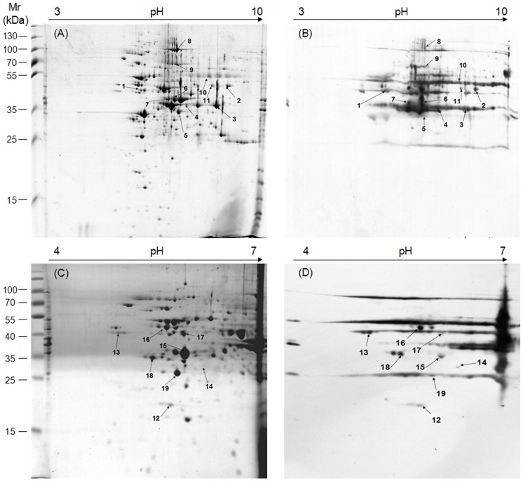
Figure 2. Two-dimensional gel electrophoresis (2-DE) and immunoblotting of the whole-cell proteins of M. bovis strain PD.
First, 350 µg and 100 µg of protein were separated by IEF using (A) a pH 3–10 IPG strip and (C) a pH 4–7 IPG strip, respectively. This was followed by SDS-PAGE on 12% gels and staining with Coomassie brilliant blue R-350. Immunoblotting was performed using each of the four antisera (Table S1) from naturally infected cattle with three replicates. The immunoreactive protein spots on PVDF membranes B and D contained all spots with good reproducibility as identified by each of the four positive sera. These corresponded to the proteins separated by 2-D gels A and C, respectively. pI values are shown on top, and the standard molecular weights are shown to the left of the gels. The spot numbers correspond to those identified by MS and listed in Table 1.

