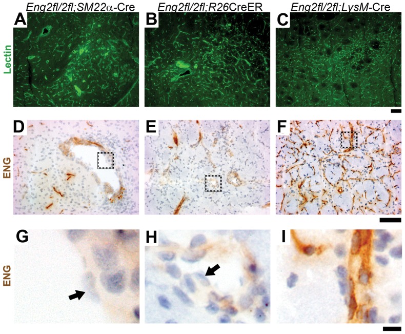
Figure 4. ENG-null endothelial cells in dysplastic vessels.
Representative images of lectin-stained brain sections from (A) AVM lesion of 5-week-old Eng2fl/2fl;SM22α-Cre, (B) VEGF-induced angiogenic focus of TM-treated adult Eng2fl/2fl;R26CreER, and (C) VEGF-stimulated angiogenic focus of adult Eng2fl/2fl;LysM-Cre mice. ENG expression in (D) Eng2fl/2fl;SM22α-Cre, (E) Eng2fl/2fl;R26CreER, and (F) Eng2fl/2fl;LysM-Cre brain. G-I: Enlarged images of the dotted boxes shown in D-F. Arrows indicate ENG-negative endothelial cells. Scale bars: 100 µm in A–F and 10 µm in G–I.

