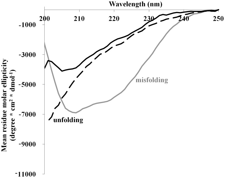
Figure 1. Trypsin structure under denaturating conditions.
0.06/ml trypsin was dissolved in buffer alone (solid black line) and in the presence of 30% TFE (gray line) or at 60°C (dashed line). In the presence of TFE, the CD spectrum showed misfolding while under heating an unfolded state was observed.

