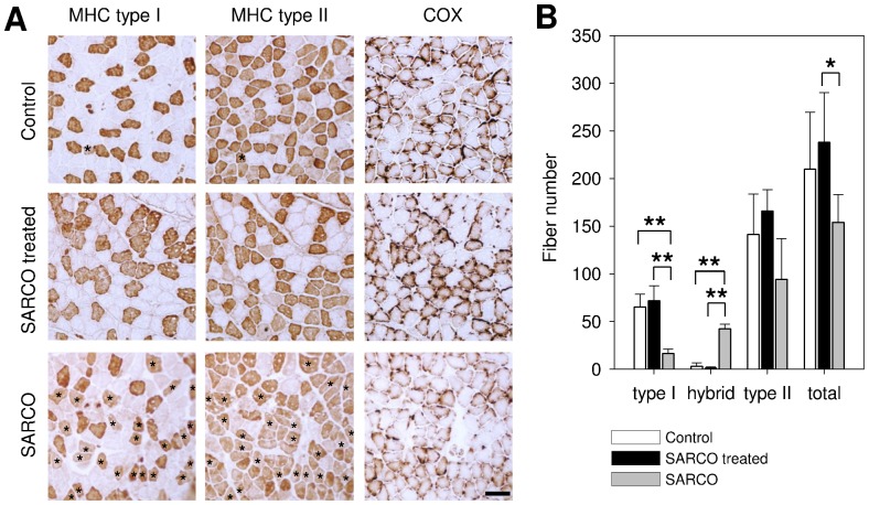
Figure 4. Fiber type distribution of m. soleus of NT-1654 and vehicle treated SARCO mice.
A: Consecutive muscle cross sections of soleus stained with myosine heavy chain (MHC) specific antibodies or cytochrome C oxidase (COX) staining as indicated. Control and treated SARCO mice show clearly separated type I and II fibers. SARCO mice have a significantly increased amount of hybrid fibers (indicated with asterisks). Cytochrome C staining of soleus sections shows a massive reduction of reactivity in SARCO mice which is reverted back to WT levels in treated SARCO mice. B: Quantitative analysis of muscle fibers of soleus. Bars represent mean values ± standard deviation. Control and treated SARCO mice have almost no hybrid fibers and are indistinguishable from each other. Compared to treated ones, SARCO mice have significantly increased hybrid fibers. Type I fibers are significantly decreased. The total fiber number in SARCO mice is also significantly decreased compared to treated animals. Number of animals: wt = 3; SARCO treated = 5, SARCO = 3. * p<0.05; ** p<0.01. Scale bar: 100 µm.

