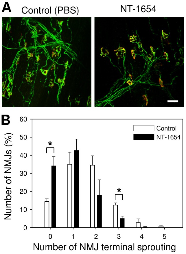
Figure 5. NMJ recovery after sciatic nerve crush.

A: Confocal images of the neuromuscular junctions (NMJs) in soleus muscle of Thy1-YFP mice after 14 days sciatic nerve crushing, treated with NT-1654 or PBS (Control) as indicated. The postsynaptic AChR was stained with Alexa-555 conjugate α-bungarotoxin (red) and the presynapse was visualized by the transgenic expression of YFP in motor neurons (green). The NMJs in soleus muscle are fully re-innervated after 14 days sciatic nerve crushing. The NMJs of NT-1654 treated mice showed significantly less nerve sprouting than those treated with PBS (Control). The number of nerve sprouting in each NMJ was counted and shown in B. NMJs in the NT-1654 treated mice have significantly fewer events of nerve sprouting and significantly fewer number of nerve sprouts than those of Control mice. Data present mean ± standard error, *: p<0.05 (two ways t-test), 100 NMJs were counted in each mouse, n = 3 mice, scale bar: 50 µm.
