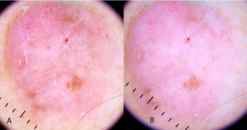
Figure 4.

Polarized (A) and non-polarized (B) dermatoscopic image of a non-pigmented lesion reveals the dermatoscopic clue of polarizing-specific white lines. Note the perpendicular orientation of these lines. This is a fibroepithelioma of Pinkus—a variant of BCC. [Copyright: ©2014 Rosendahl et al.]
