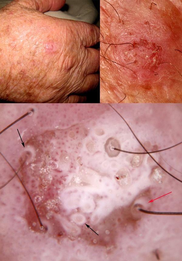
Figure 6.

Clinical and close-up images of a lesion on the hand of a 72-year-old-man which cannot be confidently diagnosed as benign clinically (upper images). Dermatoscopically (lower image) there are intensely white circles (black arrows) and one of these in particular is projected obliquely (red arrow), showing a thin white cylinder from the center of which emerges a hair shaft. White circles are produced by highly keratinized malignant squamous cells invading hair follicles. [Copyright: ©2014 Rosendahl et al.]
