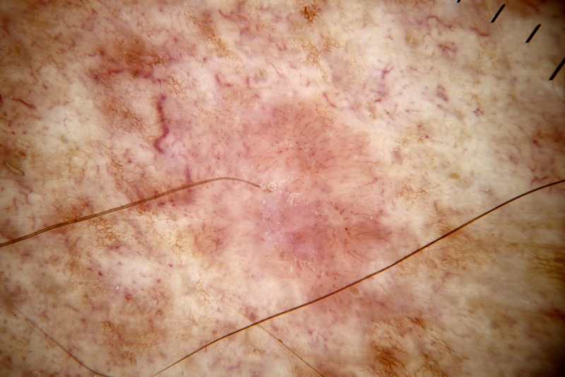
Figure 7.

Dermatoscopic image of a non-pigmented lesion. There is no ulceration and there are no “white clues” (as defined in the text). There are linear vessels covering the majority of the lesion with a pattern of dot vessels at the upper extremity. A polymorphous pattern of vessels, including a pattern of dot vessels, raises suspicion for melanoma. This was an invasive amelanotic melanoma (Breslow thickness 0.3 mm). [Copyright: ©2014 Rosendahl et al.]
