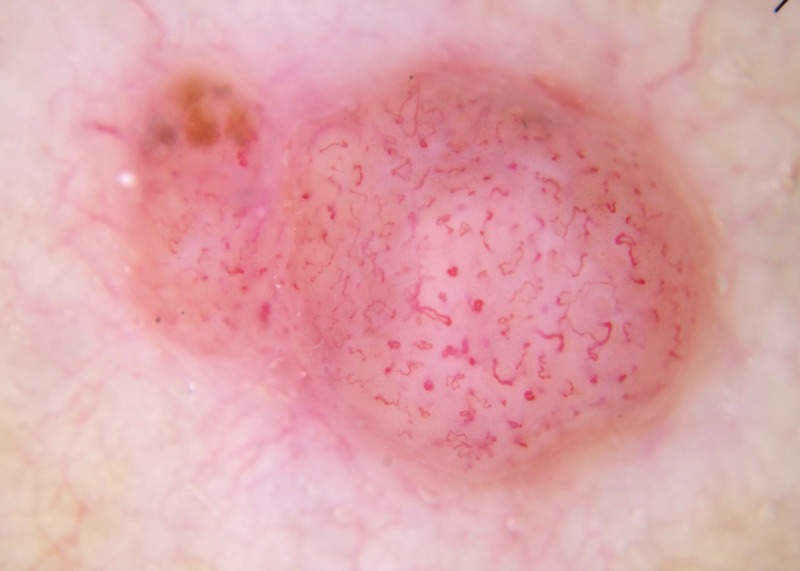
Figure 8.

Dermatoscopic image of a focally pigmented lesion. Because focal pigment is present, this should be assessed as a pigmented lesion, and as it has chaos (asymmetry of structure or color) and the clue of an eccentric structureless area, it should be excised [5]. If the focal pigment is ignored and it is assessed as a non-pigmented lesion; it is raised, the vessel pattern is non-specific and is not clods-only, so it should be excised. This was an invasive melanoma (Breslow thickness 2.5 mm). [Copyright: ©2014 Rosendahl et al.]
