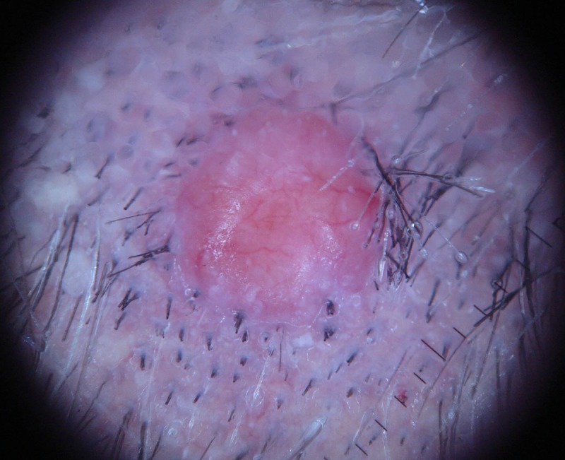
Figure 2.

Dermatoscopic view shows several scattered white globules and arborizing telangiectasia on a white to salmon pink background. The vascular branches are more pronounced at the periphery and they extend from the periphery towards the center of the lesion. [Copyright: ©2014 Cohen et al.]
