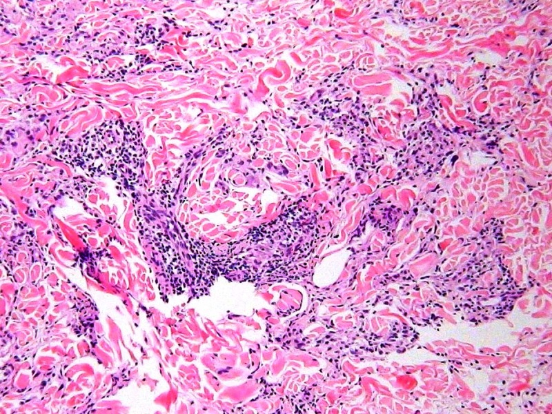
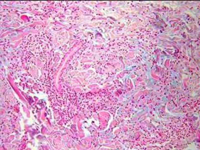
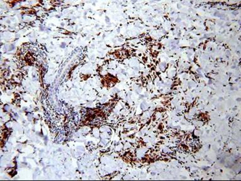
Figure 2.


Histopathological findings. (A) A high power view of hematoxylineosin stained biopsy section showing infiltrates forming a granuloma composed mainly of histiocytes and giant cells intermingled with lymphocytes. (B) Alcian-blue staining confirmed mucin deposition between collagen fibers. (C) Immunostaining for CD68 demonstrated many histiocytes and giant cells. [Copyright: ©2014 Watanabe et al.]
