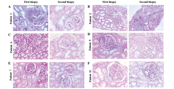
Figure 2.
Representative histological sections following immunomodulatory therapy in immunoglobulin A nephropathy. All sections were stained with periodic acid-Schiff. (A) In patient 1, at the first renal biopsy moderate diffuse mesangial cell proliferation, increased matrix deposition, adhesion to the Bowman’s capsule and interstitial mononuclear cell infiltration were observed. At the second renal biopsy, slight mesangial cell proliferation and matrix expansion, but no adhesion to the Bowman’s capsule or interstitial mononuclear cell infiltration were observed. (B) In patient 2, compared with that at the first biopsy, segmental glomerulosclerosis, interstitial infiltration, tubular atrophy and interstitial fibrosis were observed in the second renal biopsy. The histopathological features had deteriorated at the second renal biopsy (magnification, ×200). (C,D,F) In patients 4, 5 and 11, the mesangial proliferation and matrix increases observed in the first biopsy were ameliorated at the second renal biopsy. No major changes of tubulointerstitial lesions were visible (magnification, ×400). (E) In patient 7, at the first renal biopsy, glomerular cell proliferation, matrix expansion, focal adhesion to the Bowman’s capsule, tubular atrophy and interstitial fibrosis were observed. At the second renal biopsy, mild mesangial cell proliferation and adhesion to the Bowman’s capsule were detected. (A,C–F) Histopathological changes were improved at the second renal biopsy (magnification, ×400).

