
Figure 2.
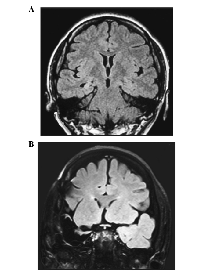
(A) Presurgical fluid-attenuated inversion recovery magnetic resonance imaging (FLAIR MRI) revealed an increased signal in the right mesial temporal structures, suggesting right mesial temporal sclerosis. (B) Postsurgical T1-weighted MRI confirmed the total removal of the right epileptogenic foci.