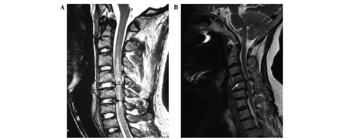
Figure 1.
Representative MRI of patients. (A) Control patient; the black arrows indicate the experimental material positions. The C4, C5 cervical vertebrae are fractured and dislocated, whereas the disc signals of C4–5, C5–6 are normal. (B) Cervical spondylosis group; white arrows indicate the surgical subtotal vertebrectomy. The discs of C4–5, C5–6 and C6–7 are herniated; the signals indicate clear disc degeneration in the cervical spondylosis group.

