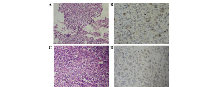
Figure 1.
Aurora-B protein expression in OS with and without pulmonary metastasis (magnification, ×400). Representative images of (A) H&E staining for OS tissues with pulmonary metastasis, showing that OS is cell rich and has significant cellular atypia, anisonucleosis, prominent nucleoli and an abundant cytoplasm; (B) IHC staining for Aurora-B protein with lung metastasis, showing brown-yellow particles deposited in the nucleus and coloring of the majority of the cells; (C) H&E staining for OS tissues without pulmonary metastasis, showing that OS is cell-rich and has significant cellular atypia, anisonucleosis, prominent nucleoli, an abundant cytoplasm and a small quantity of bone-like matrix; (D) IHC staining for Aurora-B protein in OS tissues without pulmonary metastasis, showing brown-yellow particle deposition in the nucleus and coloring of only a few cells. OS, osteosarcoma; H&E, hematoxylin and eosin; IHC, immunohistochemistry.

