Figure 5.

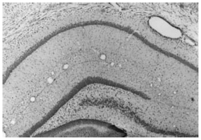
Nissl staining in the sham-surgery group. The neurons in the cerebral cortex and hippocampus were densely distributed (magnification, ×100).

Nissl staining in the sham-surgery group. The neurons in the cerebral cortex and hippocampus were densely distributed (magnification, ×100).