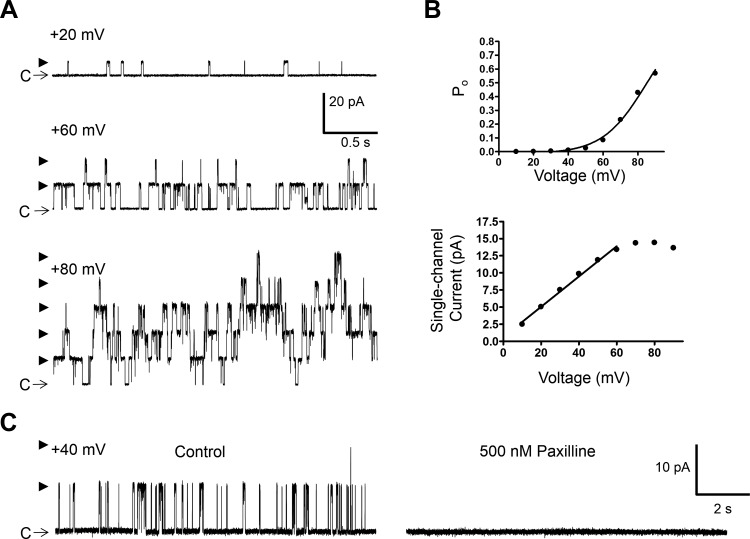
Fig. 2.
Excised patch single BK channel recording from a freshly isolated native DSM cell in the presence of low intracellular Ca2+ concentration ([Ca2+]). A: representative single BK channel currents measured at the indicated voltages demonstrating an increase in channel activity with depolarization. C depicts the closed channel state and arrowheads the open channel states. B: single-channel current-voltage and open probability (Po)-voltage graphs for the patch represented by traces in A. C: single BK channel activity measured at +40 mV before and after the addition of paxilline (500 nM) showing inhibition of the BK channel opening. Data and recordings in A–C were obtained from the same inside-out patch using symmetrical 140 mM K+ solution with ∼300 nM free [Ca2+].

